Neuronal loss, a pathological hallmark of Alzheimer’s disease, is recapitulated in reMYND’s transgenic human hTauP301L mouse model. Immunohistochemical staining and quantification of NeuN positive neuronal nuclei of 9 month old hTauP301L mice demonstrate a 20 % neuronal loss in the subthalamic nucleus in comparison to age- and sex-matched control mice and the younger hTau.P301L mice (5 months) (Figure 1).
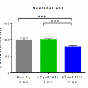
Figure 1: anti-NeuN in the subthalamic nucleus - zona incerta. Quantifications are shown as % reactive area of the region of interest and relative to the NeuN reactive area in non-trangenic controls (100%). * p < 0.05, ** p < 0.01, *** p < 0.001, **** p < 0.0001. Non-Tg: non-transgenic control mice
In comparison to the 5 months old hTauP301L mice and wild type controls, the aged hTauP301L mice show a prominent neurofibrillary tangle pathology (Figure 2, AT8 and AT100) indicating that the Tau pathology is steered by transgenic expression of human TauP301L drives neuronal loss
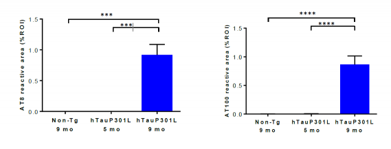
Figure 2: AT8 (pSer202 and pThr205, left) and AT100 (pThr212 and pSer214, right) immunohistological staining in the interposed cerebellar nucleus, anterior and posterior part (deep cerebellar nuclei). Quantifications are shown as % reactive area of the region of interest. * p < 0.05, ** p < 0.01, *** p < 0.001, **** p < 0.0001.
For more information on reMYND’s transgenic mouse models or read-outs, please visit our website or contact Bart Roucourt, CRO Manager.
With kind regards,
The CRO team
reMYND, Bio-Incubator (Wetenschapspark)
Gaston Geenslaan 1
BE-3001 Leuven (Heverlee)
Belgium
RPR Leuven, 0476.910.101
BTW/VAT: BE0476.910.101
T +32 16 75 14 20
F +32 16 75 14 21
info@remynd.com