Two-photon imaging allows tracking the formation of Tau aggregates in the same brain-region of a live animal over the course of time. This makes it possible to study the effects of treatment longitudinally in vivo. In addition to studying compounds’ efficacy on NFT-formation, this high resolution imaging technique also allows for advanced PK/PD studies with labelled antibodies in the cortex of Tau.P301S animals. In the course of such studies, it is possible to determine the exact location of the antibody relative to the location of Tau aggregates as well as to quantify the antibody turn-over.
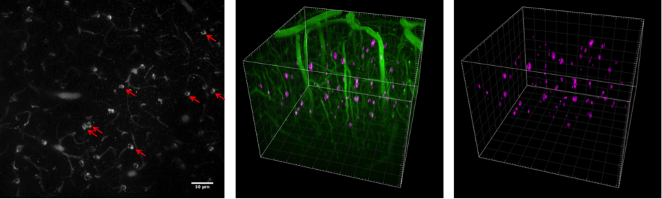
Figure 1: in vivo two-photon images of superficial cortex layers in a 5.5 month old Tau.P301S animal 24h following Methoxy-X04 injection (ip) are shown as an optical section (left) and as a 3D projection (middle and right). Labelled Tau aggregates are indicated with red arrows (left) or highlighted using purple pseudo-color (middle and right). Scalebar and grid: 50 μm.
The Tau.P301S model features progressive hyperphosphorylation of Tau with ageing (Figure 2), which leads to a profound NFT-like pathology in the brainstem, spinal cord, cortex and hippocampus regions and to impaired Long Term Potentiation at 3 months of age.

For more information on reMYND’s transgenic mouse models, please contact Bart Roucourt, CRO Manager.
reMYND, Bio-Incubator (Wetenschapspark)
Gaston Geenslaan 1
BE-3001 Leuven (Heverlee)
Belgium
RPR Leuven, 0476.910.101
BTW/VAT: BE0476.910.101
T +32 16 75 14 20
F +32 16 75 14 21
info@remynd.com