Key characteristics
- β-amyloid plaques are observed in the cortex, hippocampus and subiculum from an age of 8 months
- Tau pathology is observed in the cortex, hippocampus, subiculum and brainstem from an age of 8 months
- Astrocytosis and microgliosis are observed in the cortex and subiculum from an age of 9 months
- Cognitive impairment in Morris water maze paradigm from an age of 6.5 months
- Progressive motoric impairment from an age of 9 months

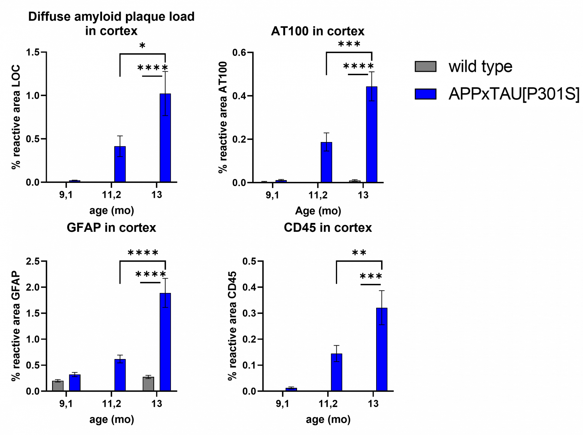
Progressive accumulation of plaque deposition (LOC), hyperphosphorylated Tau (AT100 = pThr212/pSer214 Tau), astrocytosis (GFAP) and microgliosis (CD45) in the cortex as measured by IHC (mean ± SEM, n = 8, no data for WT at 11.2 months). Statistics: One-Way ANOVA, Tukey’s multiple comparison.

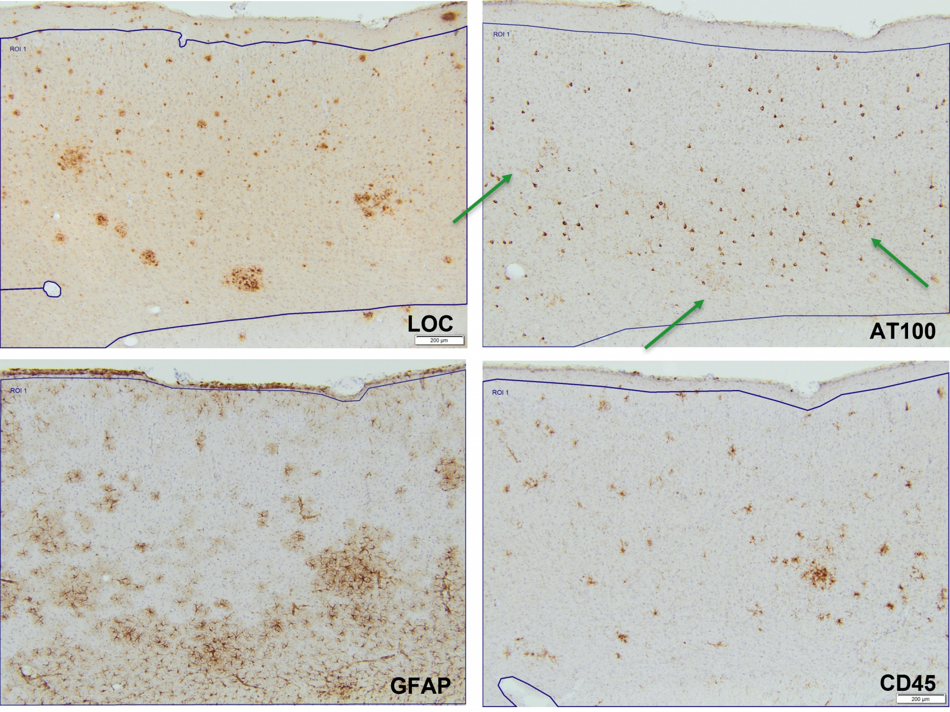
Adjacent brain section of a single mouse at 13 months of age of Aβ, Tau and inflammatory markers.

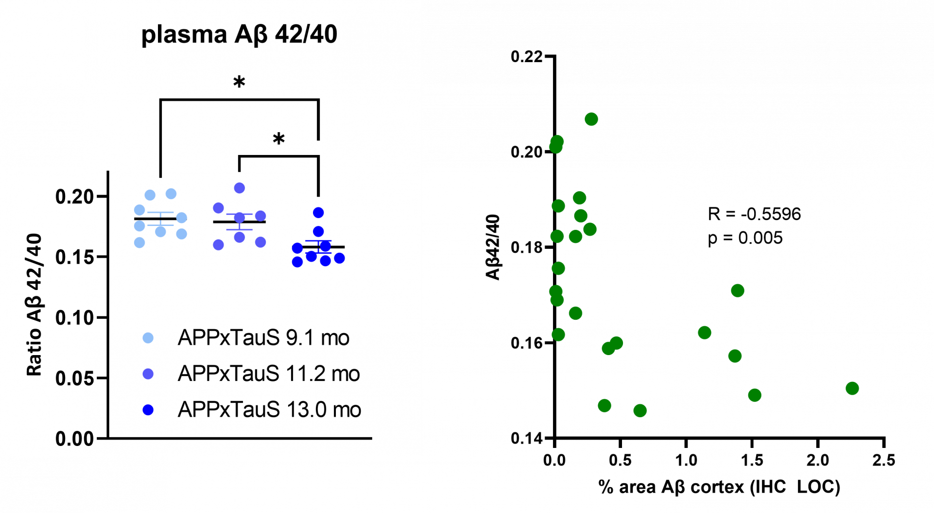
Progressively lower Aβ42/40 in plasma correlates with Aβ plaques in the cortex (mean ± SEM, n = 7 or 8). Plasma Aβ concentration were measured using MSD-technology (kit K15200E). A significant negative correlation is observed between the plasma Aβ42/40 ratio and Aβ pathology in the cortex. Statistics: One-Way ANOVA, Tukey’s multiple comparison and Pearson’s r correlation coefficient.
Interested to see more data?
We have a more extensive datapackage available. Contact us at cro[at]remynd.com to set up a meeting and discuss how we translate your mechanism of action into an effective study design.


